Malignant melanoma is a serious type of skin cancer which occurs due to uncontrolled growth of pigment cells called melanocytes.
Over a 10-year period study in Malta between 1993 and 2002, 13% of the total number of malignant melanomas occurred in people under 35 years of age, 47% in 36-60 year-olds, and 40% in those over 61years of age.
The good news is however that, if detected early enough, most thin melanomas are fully cured. One should therefore routinely examine his or her skin and note whether any new moles are forming or whether existing moles have changed colour or shape. If a number of moles are present, it is advisable to have these moles photographically mapped by a qualified medical photography practitioner. This will help you and your dermatologist to determine whether there were any changes in colour, shape or size in any one of the moles from your last visit. This is the main concept of whole body mole mapping.
MOLE MAPPING
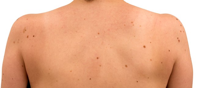
Mole Mapping is a procedure whereby your moles are catalogued or “mapped”. Mole mapping provides a set of around 20 high quality, standardized photographs of the whole body. These photos will document all the existing moles and will help the dermatologist to keep a visual track of your moles hence assisting him or her in the early diagnosis of malignant melanoma. Having a set of photographs to monitor existing moles could be a life saver. Mole mapping is a specialised photographic process, undertaken only by qualified medical photography practitioners with no intervention.
